Microdiscectomy
A discectomy refers to the excision of the intervertebral disc material that may be described as herniated, implying that it is “bulging” or “ruptured” through the ligaments. If the central fragment of disc material has torn through a hole in the ligament, it is called an extruded fragment or extruded disc. The term herniated nucleus pulposus (HNP) is a catchall phrase for all of these conditions. The prefix “micro” is used to denote that the operation is done through use of an operating microscope where Dr. Kim will employ microsurgical techniques.
Generally, a discectomy is under the umbrella of a decompression type procedure. This procedure is carried out to relieve pressure on the spinal cord or nerve roots. The pressure may result from fracture fragments, disc fragments, bone spurs, tumors or infections.
Your back and leg symptoms are caused by pressure on a nerve. This pressure comes from a damaged disc or by bone growth. Constant wear and tear can make a disc weak and worn out. This allows the disc to bulge. If the outside of the disc (the annulus) tears the soft insides of the disc can herniate out. This can pinch your nerves. As your spine ages, bone spurs can form and grow into the foramen (area where the nerve comes out) of your vertebrae. The bone spurs then press on your nerves in the spinal canal or through the foramen. The joints in your spine (facets) can enlarge with arthritis and press on your nerves. The ligament that holds your vertebra together may also thicken causing further spinal stenosis (narrowing of the spinal canal) and pressure on the nerves.
A minimally invasive discectomy and/or decompression (fig 1 and 2) is done through a small incision using a tubular retractor that measures between 14mm and 18mm. This leads to an incision about the size of a nickel or a quarter depending on how many levels need to be decompressed. Typically, this spine surgery is done on an ambulatory basis where the patient leaves the same day following surgery.
Lumbar Microdiscectomy
Surgery often helps to relieve your leg pain, numbness, tingling and weakness. This is done by eliminating the pressure on the nerves by removing disc material or bone from the spine. This will allow you to move with greater ease.
Dr. Kim will do a surgery to relieve this pressure. He will make an incision on your back that will allow him to access the affected area. The incision size will vary depending on how many levels of your spine Dr. Kim needs to address. Dr. Kim uses a microscope during the surgery to help enlarge the view of your spine and ensure accuracy. Dr. Kim will remove a small piece of bone (the lamina) to allow access to the herniated disc, ligament, or bone spurs that may be pressing on your nerves. This is called a laminotomy. If there is no herniated disc, the laminotomy alone will help take pressure off of the nerve. He may also remove bone in the foramen area, if necessary (foraminotomy). This will allow room for your nerves to breathe and take the pressure off of them, thus eliminating pain, weakness, and numbness.
When the surgery is completed, Dr. Kim will close your incision with dissolvable sutures under the skin and DERMABOND® PRINEO® on the outside of your incision.
Post-operative pain is different for each patient. Numbness is always the last thing to improve after this surgery. It can take twice as long as you have had the numbness for sensation to return. For example, if you had a numb left foot for one month, it may take two months to show improvement of the numbness. Pain and strength respond very quickly. For the first six weeks, some patients experience intermittent radiating pain similar to the pain they had before surgery. This is related to the fact that Dr. Kim had to protect the nerve during surgery. It can also be inflammation from having surgery and nerve recovery. This pain should subside with time. Please contact Dr. Kim’s office with any complaints of consistent pain or if you have any questions about your post-operative pain.
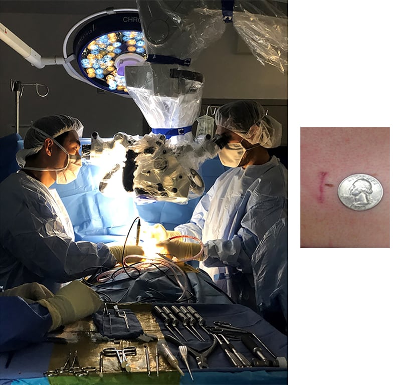
Fig 1a A surgical microscope is used to perform a minimally invasive operation.
1b The incision size may vary depending on the number of levels that need to be decompressed. Typically the Incision is between 1.5 and 2.5cm (about the diameter of a nickel or quarter).
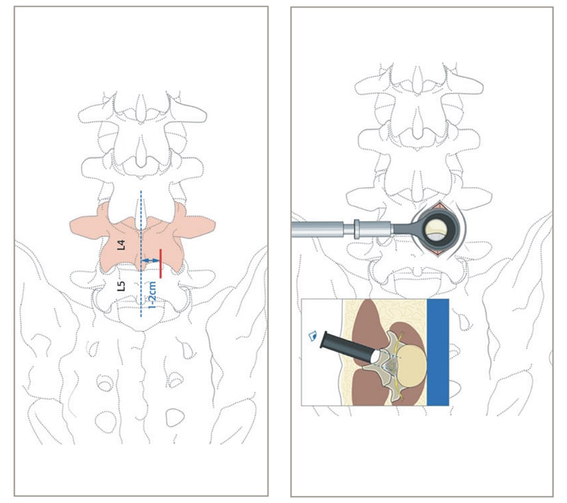
1c A minimally invasive discectomy and decompression may be indicated in a patient such as one shown in figure 1c. A disc herniation at L4-5 causing symptoms of “sciatica” or nerve pain running down the leg.
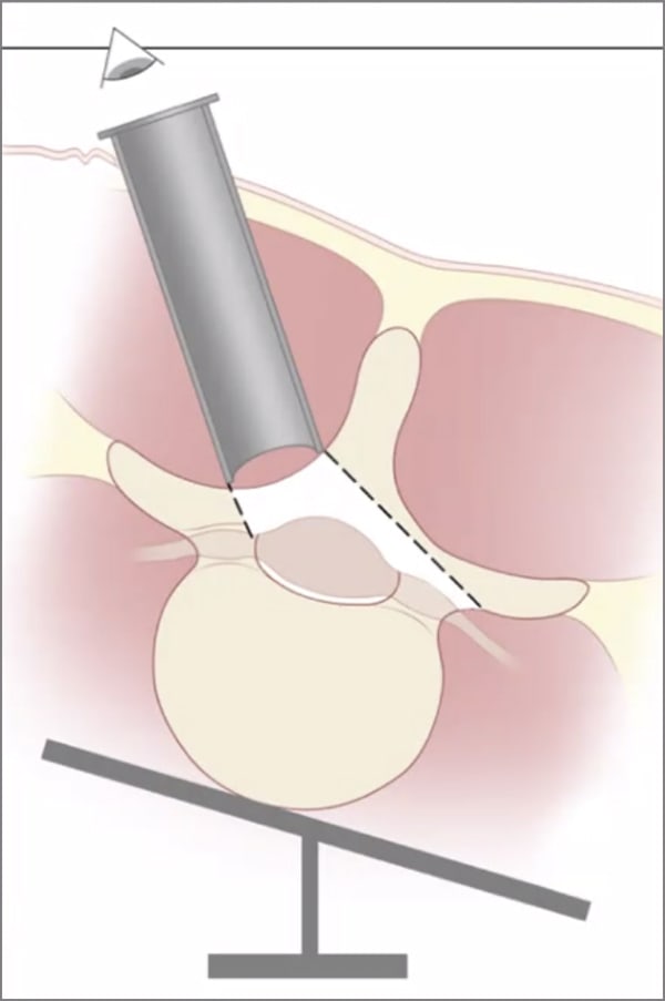
Fig 2 showing a minimally invasive discectomy and approach. Note that the tubular retractor that Dr. Kim uses measures between 14mm-18mm for a typical microdiscectomy.